


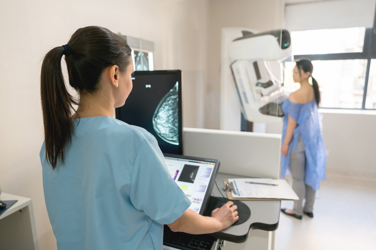
Ductal intracellular carcinoma (DCIS) is a kind of pre-invasive tumor that may progress to one of the deadly types of breast most cancers. It accounts for about 25 p.c of all breast most cancers diagnoses.
As a result of it’s troublesome for clinicians to find out the sort and stage of DCIS, DCIS sufferers are sometimes overtreated. To handle this challenge, an interdisciplinary crew of researchers from MIT and ETH Zurich developed an AI mannequin that may establish completely different phases of DCIS from cheap, available breast tissue photographs. The mannequin exhibits that each the state and association of cells inside a tissue pattern are essential in figuring out the stage of DCIS.
As a result of such tissue photographs are available, the researchers constructed one of many largest datasets of its sort, utilizing it to coach and take a look at their mannequin, and in contrast its predictions with the conclusions of pathologists, usually with clear settlement.
Sooner or later, this mannequin may very well be used as a device to assist clinicians extra effectively diagnose less complicated circumstances with out the necessity for labor-intensive testing, permitting extra time to judge circumstances the place it’s unclear whether or not DCIS will grow to be invasive.
“We have taken the primary steps in understanding that it is advisable to have a look at the spatial group of cells to diagnose DCIS, and now we’ve got developed a scalable know-how. From right here, we actually want forward-looking analysis. Working with hospitals and bringing this into the clinic shall be a key step ahead,” says Caroline Uhler, professor within the Division of Electrical Engineering and Pc Science (EECS) and the Institute for Information, Methods, and Society (IDSS), director of the Eric and Wendy Schmidt Middle on the Broad Institute of MIT and Harvard, and a researcher in MIT’s Institute for Info and Resolution Methods (LIDS).
Uhler co-authored the research, together with lead creator Xinyi Zhang, a graduate scholar at EECS and the Eric and Wendy Schmidt Middle, and co-author GV Shivashankar, ETH Zurich professor of mecogenomics in collaboration with the Paul Scherrer Institute, in addition to different researchers from MIT, ETH Zurich and the College of Palermo in Italy. The open entry research is Released on July 20th Nature Communications.
Combining Imaging and AI
Thirty to 50 p.c of DCIS sufferers develop extremely invasive stage most cancers, however researchers do not know the biomarkers that might inform clinicians which tumors will progress.
Researchers can use strategies reminiscent of multiplex staining and single-cell RNA sequencing to find out the stage of DCIS in tissue samples, however these assessments are too costly to be extensively accessible, Shivashankar explains.
In earlier research, the researchers have proven that a cheap imaging method known as chromatin staining might be as informative because the rather more costly single-cell RNA-sequencing evaluation.
The research hypothesized that this single stain, mixed with a rigorously designed machine studying mannequin, might probably present the identical details about the stage of most cancers as costlier strategies.
First, the analysis crew created a dataset containing 560 photographs of tissue samples from 122 sufferers at three completely different phases of the illness. They used this dataset to coach an AI mannequin that discovered representations of the state of every cell within the tissue pattern photographs to deduce the stage of a affected person’s most cancers.
However not all cells present indicators of most cancers, so the researchers wanted to combination them in a significant manner.
The researchers designed a mannequin to create clusters of cells with related states and recognized eight states which can be key markers for DCIS. Some cell states are extra indicative of invasive most cancers than others. The mannequin determines the share of cells of every state in a tissue pattern.
Group issues
“However in most cancers, the composition of cells additionally adjustments. We discovered that it is not sufficient to simply know the share of cells in each state; we additionally want to grasp how the cells are composed,” says Shivashankar.
Armed with this perception, the researchers designed a mannequin that took under consideration the proportions and association of cell states, considerably bettering accuracy.
“What was attention-grabbing for us was to see how essential the spatial group is. Earlier research have proven that cells near the ducts are essential. However it’s additionally essential to think about which cells are near which different cells,” Zhang says.
Once they in contrast the mannequin’s outcomes with samples evaluated by pathologists, they discovered a transparent settlement in lots of circumstances. In much less clear circumstances, the mannequin additionally supplied details about the tissue pattern’s traits, reminiscent of mobile composition, that pathologists might use to tell their decision-making.
This versatile mannequin may be relevant to different forms of most cancers and neurodegenerative illnesses, an space the researchers are at the moment investigating.
“We have proven that with the precise AI strategies, this straightforward staining might be very highly effective. There’s nonetheless numerous analysis to do, however extra analysis must keep in mind mobile group,” Uhler says.
This analysis was funded by the Eric and Wendy Schmidt Middle on the Broad Institute, ETH Zurich, the Paul Scherrer Institute, the Swiss Nationwide Science Basis, the U.S. Nationwide Institutes of Well being, the U.S. Workplace of Naval Analysis, the MIT Jameel Machine Studying and Well being Clinic, the MIT-IBM Watson AI Lab, and a Simons Investigator Award.
